
Case32
●1979
Age at initial visit: 8 years and 7 months; female / Protruding lower bite., Deep bite
Treatment duration:Phase I treatment: 1 y 1 m
Phase II treatment: 1 y 9 m
No extractions
Age at initial visit: 8 years and 7 months; female / Protruding lower bite., Deep bite
Treatment duration:Phase I treatment: 1 y 1 m
Phase II treatment: 1 y 9 m
No extractions

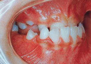
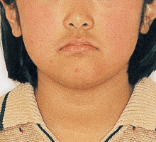
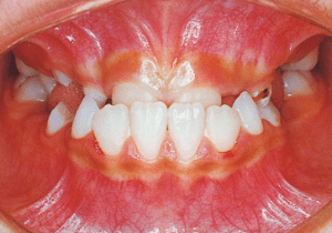
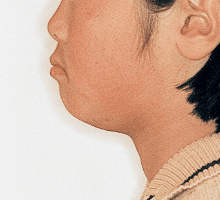
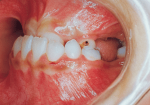
1 Initial Visit 3-10-’79
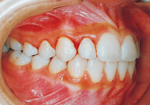
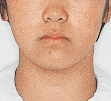
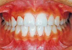
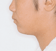
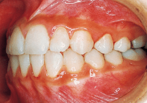
2 After Phase 2 Treatment 10-11-’83
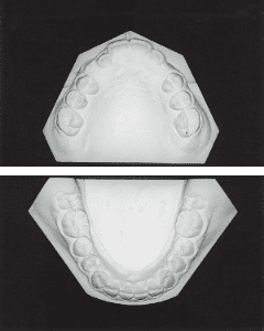
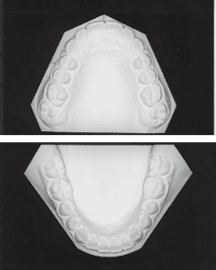
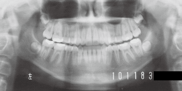
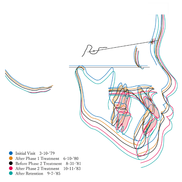
Protruding lower bite during the mixed dentition phase. A case in which the maxilla was expanded during Phase I and Phase II was completed without extractions. The primary goal of Phase I treatment was to address the relatively narrow maxillary alveolar bone—which was determined to have been stunted due to the early loss of primary teeth—by expanding it to improve the overjet of the anterior teeth. Based on the condition of the lower jaw’s dentition and the skeletal structure, the Phase I treatment plan was designed with the goal of achieving non-extraction in Phase II.
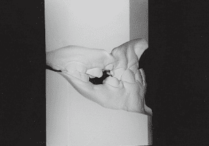
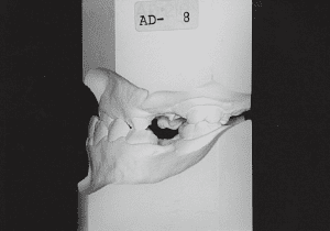
3 Initial Visit 3-10-’79
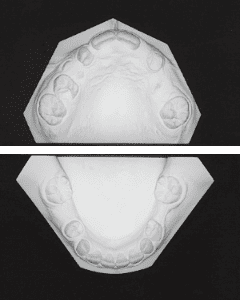
3 Initial Visit 3-10-’79
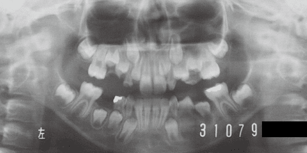
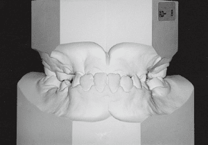
4 Initial Visit 3-10-’79
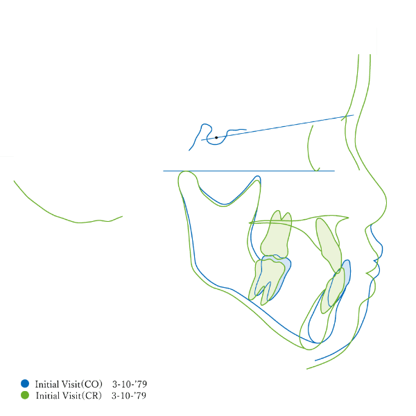
5
Although the model shows a Class I underbite, the maxillary arch is significantly smaller than the mandibular arch. There are many missing primary teeth, and dental arch disruption is particularly severe in the maxilla (3). When closing the mouth, contact between the anterior teeth causes the mandible to be guided slightly forward. Skeletal disharmony is not as pronounced as the malocclusion suggests (5); it was determined that this was the result of the strong morphological effects of early loss of deciduous teeth.
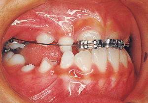
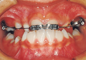
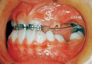
6 4-10-’79 The beginning of first phase 8 years and 8 months old
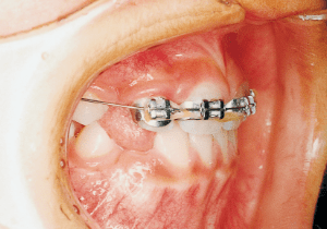
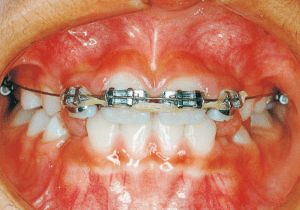
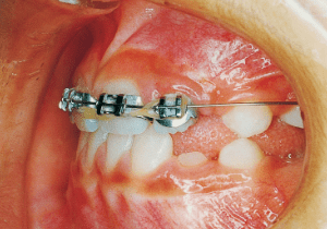
7 4-8-’80 During observation 9 years and 8 months old
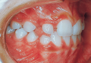
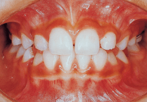
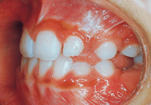
8 6-10-’80 After Phase 1 Treatment 9 years and 10 months old
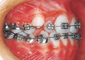
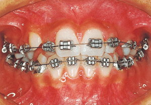
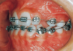
9 10-28-’81 Before Phase 2 Treatment 11 years and 2 months old
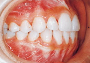
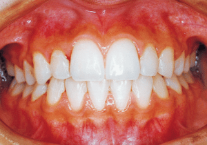
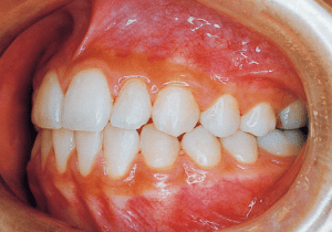
10 9-7-’85 After Retention 15 years and 1 months old
In the Phase I treatment, appliances were placed only on the maxilla. No intermaxillary or extraoral anchorage was used (6). The molar relationship was finalized as Class I, and space was created to improve the overjet of the anterior teeth and accommodate the eruption of permanent teeth (7). This condition was maintained for a period of time, and the appliances were removed (8). As canine eruption began, treatment with full braces was initiated. Figure 10 shows the occlusion at the conclusion of retention.
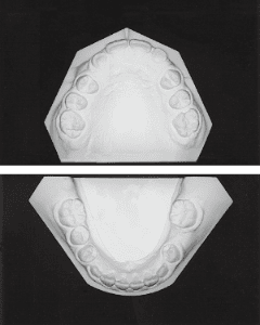
11 After Phase 1 Treatment 6-10-’80
12 Before Phase 2 Treatment 8-31-’81
13 After treatment 10-11-’83
14 After treatment 10-11-’83
15